Jerit Mitchell stared at his computer screen in disbelief. The undergraduate physics student had been analyzing X-ray scans of a 66-million-year-old dinosaur bone when something impossible appeared in the data.
Twisted, branching structures snaked through the ancient rib bone like underground rivers. His mentors gathered around the monitor, pointing at formations that shouldn’t exist in fossilized remains. What they were seeing challenged everything science textbooks taught about dinosaur preservation.
Soft tissues disappear within years after death. Everyone knows that. Bones and teeth survive because they’re made of minerals, but blood vessels, muscles, and organs vanish completely. Yet here was evidence of something that defied paleontology’s fundamental rules.
Six years later, Mitchell’s discovery has rewritten scientific understanding of what’s possible in the fossil record. The finding opens doors to studying dinosaur biology in ways researchers never imagined.
Student Makes Discovery That Rewrites Dinosaur Science

Mitchell’s 2019 discovery occurred while he was reviewing routine scans at the University of Regina in Canada. As an undergraduate physics student working with particle accelerators to study fossils, he possessed the technical skills to spot anomalies others might miss.
Initial analysis suggested equipment malfunction or scanning artifacts. Sophisticated synchrotron X-ray machines sometimes produce strange images, and student researchers learn to distinguish real data from technical errors.
However, repeated scans confirmed the structures were genuinely embedded within the bone tissue. Multiple imaging techniques revealed the same branching, vessel-like formations throughout the specimen.
Mitchell’s physics background provided a crucial perspective for interpreting the unusual findings. His interdisciplinary approach combined advanced imaging technology with paleontological expertise from seasoned researchers.
The discovery launched years of detailed analysis that would eventually earn publication in Scientific Reports, one of science’s most prestigious journals.
Meet Scotty: The Largest T. Rex Ever Found
The blood vessels came from an exceptional Tyrannosaurus rex specimen nicknamed Scotty, discovered in Saskatchewan, Canada, in 1991. Paleontologists celebrated the find by drinking Scotch whisky, giving the dinosaur its memorable name.
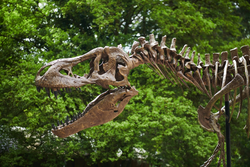
Scotty represents the largest T. rex ever found, estimated to have weighed nearly 20,000 pounds during life. The massive predator measured over 40 feet long and stood more than 12 feet tall at the hip.
Sixty-five percent of Scotty’s skeleton was recovered from the Late Cretaceous Frenchman Formation, making it one of the most complete T. rex specimens ever discovered. The exceptional preservation enabled detailed studies of dinosaur anatomy and pathology.
Evidence from Scotty’s bones suggests the giant predator lived a violent life filled with combat injuries, diseases, and trauma. Bite marks, fractures, and other damage tell stories of prehistoric battles fought 66 million years ago.
Today, a replica of Scotty’s skeleton amazes visitors at the Royal Saskatchewan Museum while the original bones continue revealing scientific secrets in research laboratories.
When X-rays Revealed the Impossible

Synchrotron X-ray imaging technology made Mitchell’s discovery possible by peering inside fossil bones without causing damage. These high-intensity X-rays, produced at specialized particle accelerator facilities, penetrate dense mineralized tissues with unprecedented clarity.
Standard medical CT scanners lack sufficient power to examine fossilized dinosaur bones. Mineralization during fossilization creates extremely dense structures that ordinary imaging equipment cannot penetrate effectively.
3D modeling software converted the X-ray data into detailed visualizations of internal bone structure. Layer by layer, the scans revealed branching networks of preserved soft tissues hidden within Scotty’s rib.
Non-invasive analysis protects irreplaceable fossil specimens from damage during scientific study. Traditional paleontological techniques required cutting or grinding bones to examine internal structures, often destroying evidence in the process.
Digital reconstruction allowed researchers to examine the preserved blood vessels from multiple angles and create physical models using 3D printing technology.
Blood Vessels That Shouldn’t Exist After 66 Million Years
The discovery challenged fundamental assumptions about fossil preservation that scientists had held for centuries. Mitchell explained the significance: “Normally, what gets preserved in the fossil record is only just the hard parts—just the bones or the teeth. But we can actually have the soft tissues preserved in rare circumstances, and these can tell us a lot more about how dinosaurs lived millions of years ago.”
Organic tissues typically decompose completely within years or decades after death. Bacteria, environmental conditions, and chemical processes break down proteins, blood vessels, muscles, and other soft structures long before fossilization occurs.
Exceptional preservation requires specific environmental conditions that prevent normal decay processes. Rapid burial, oxygen-free environments, and particular chemical compositions can sometimes preserve soft tissues for extraordinary lengths of time.
Previous discoveries of dinosaur soft tissues sparked heated scientific debates about authenticity and preservation mechanisms. Many researchers remained skeptical that original biological materials could survive geological timescales.
Scotty’s blood vessels provide new evidence that soft tissue preservation occurs more frequently than previously believed, opening possibilities for studying dinosaur biology in unprecedented detail.
Scotty’s Battle Scars Tell a Violent Story

The preserved blood vessels appeared specifically within a partially healed rib fracture, providing clues about Scotty’s violent final months. Paleontologists have documented numerous injuries across the skeleton, suggesting this T. rex survived multiple combat encounters.
Bite marks on various bones indicate attacks from other large predators, possibly during territorial disputes or fights over food sources. Tyrannosaur combat behavior likely resembled modern crocodile fighting, with powerful jaws inflicting devastating injuries.
Evidence suggests Scotty’s rib fracture was still healing when death occurred, indicating survival for several months after the initial injury. Incomplete bone repair processes created conditions that enabled exceptional soft tissue preservation.
Adult male T. rex specimens commonly show combat-related injuries, supporting theories about aggressive territorial behavior and dominance battles. Successful predators needed to defend hunting grounds and mating territories from competitors.
The healing fracture reveals T. rex possessed robust recovery capabilities, allowing individuals to survive serious injuries that might prove fatal to other animals.
How Healing Bones Created Perfect Preservation
Co-author Mauricio Barbi explained the preservation mechanism: “Preserved blood vessel structures, like we have found in Scotty’s rib bone, appear linked to areas where the bone was healing. This is because during the healing process, those areas had increased blood flow to them.”
Angiogenesis, the formation of new blood vessels, occurs during bone repair as the body rushes nutrients and immune cells to injury sites. Fractured bones develop extensive vascular networks to support healing processes.
Enhanced blood flow during healing created ideal conditions for the fossilization of vascular structures. Iron-rich blood provided mineral content that replaced organic tissues while preserving their three-dimensional architecture.
The timing of Scotty’s death during active healing explains why these particular blood vessels survived fossilization. Established bone tissue lacks the dense vascular networks found in healing injuries.
Researchers now target pathological bones showing signs of injury or disease when searching for preserved soft tissues, increasing the chances of making similar discoveries.
Iron-Rich Secrets Inside Ancient Bones

Chemical analysis revealed the blood vessels had been preserved as iron-rich mineralized casts composed primarily of pyrite, goethite, and hematite. These iron compounds replaced the original organic tissues while maintaining their structural shape.
Two distinct layers of mineralization occurred during preservation, reflecting complex environmental changes in the burial site over millions of years. The first layer consisted of fine-grained pyrite, while the second featured larger crystalline structures.
Iron content in the vessels came from hemoglobin in the original blood, providing the chemical foundation for mineralized preservation. As organic compounds decayed, iron minerals precipitated in their place.
Manganese and other trace elements also contributed to the preservation process, creating chemical signatures that distinguish preserved tissues from the surrounding rock matrix. Synchrotron analysis detected these subtle compositional differences.
The specific preservation chemistry suggests particular environmental conditions existed at the burial site, including low-oxygen conditions and iron-rich groundwater that enabled exceptional fossilization.
Particle Accelerators Unlock Dinosaur Mysteries
Advanced synchrotron facilities like the Canadian Light Source provide paleontologists with powerful tools for non-destructive fossil analysis. High-energy X-rays penetrate dense fossil bones while revealing microscopic internal structures.
Particle accelerator technology produces X-rays thousands of times more intense than conventional medical scanners. This extraordinary power enables imaging of heavily mineralized fossils that would be invisible to standard equipment.
3D reconstruction techniques convert raw X-ray data into detailed three-dimensional models that scientists can manipulate and study from any angle. Digital analysis reveals structural details impossible to see with traditional methods.
Multiple imaging techniques applied to the same specimen provide different types of information about chemical composition, mineral structure, and tissue organization. Combined analysis creates detailed pictures of ancient biology.
Synchrotron facilities worldwide are revolutionizing paleontology by enabling discoveries that seemed impossible just decades ago, opening new research directions in the study of extinct life.
Rewriting the Rules of Fossilization

Jordan Mallon, a paleontologist at the Canadian Museum of Nature not involved in the research, emphasized the discovery’s broader significance: “For centuries, it’s been thought that there’s effectively no trace of biological tissue in a fossil—that there shouldn’t be. And yet, as we start to put these things under the microscope and look at them with new techniques, and look at them in more depth, it turns out the fossilization process isn’t quite as straightforward—or maybe not as rapid—as we thought it would be.”
Traditional paleontology focused primarily on hard tissues like bones and teeth, assuming soft structures never survived geological timescales. Recent discoveries challenge these assumptions with mounting evidence of preserved proteins, blood vessels, and other organic remains.
Exceptional preservation requires specific environmental conditions, including rapid burial, chemical stability, and protection from bacterial decomposition. These conditions occur more frequently than previously believed.
Modern analytical techniques reveal preservation evidence that earlier generations of scientists lacked the tools to detect. Advanced microscopy, chemical analysis, and imaging technology unlock information hidden within fossils.
The expanding field of molecular paleontology promises even more surprising discoveries as technology continues to advance and researchers develop new analytical approaches.
The Future of Impossible Discoveries
Mitchell’s breakthrough demonstrates how interdisciplinary research combining physics, chemistry, and paleontology can yield revolutionary insights about ancient life. Cross-disciplinary collaboration accelerates scientific progress by bringing diverse expertise to complex problems.
Future research will target additional pathological dinosaur bones showing signs of injury, disease, or healing to search for more preserved soft tissues. Success rates should improve as researchers learn to identify promising specimens.
Technology improvements continue to expand possibilities for fossil analysis. New imaging techniques, chemical methods, and analytical tools may reveal preservation evidence currently beyond detection limits.
The discovery encourages researchers to revisit museum collections with fresh perspectives and modern analytical capabilities. Specimens collected decades ago may harbor secrets that contemporary technology can finally unlock.
As this field develops, scientists may eventually recover enough preserved soft tissues to reconstruct detailed pictures of dinosaur physiology, behavior, and ecology that seemed impossible just years ago.
