A frantic late-night Google search and a mother’s refusal to accept an uncertain answer helped save a six-year-old boy’s life after doctors initially diagnosed him with the flu. What looked at first like a routine childhood illness spiralled into a medical emergency so severe the youngster lost the ability to walk, talk and breathe on his own — until a specialist’s intervention reversed the course.
This is the story of Witten Daniel and his mother, Casey Daniel — a family from Lubbock, Texas — and how a combination of instinct, online research and rapid transfer to a specialist team in Houston gave the boy a second chance.
A Rapid Decline: When “It’s Just the Flu” Wasn’t Enough
In April, Witten started with what seemed like common flu symptoms: dizziness, headache and fatigue. Local doctors initially treated him for influenza, but within hours his condition deteriorated sharply. According to local reporting, he lost the ability to walk, talk and even breathe independently and soon required intubation and life support in the hospital.
The speed of the decline alarmed his mother, Casey. What had begun as fever and malaise quickly turned into what looked like a neurological emergency — a pattern that didn’t fit an ordinary case of flu.

How One Google Search Changed the Course of Care
Refusing to accept that there was nothing more to be done, Casey began searching symptoms on her phone. Her queries led her to the work of a neurosurgeon who specializes in the kind of lesions the doctors were worried about. Casey reached out to Dr. Jacques Morcos at UTHealth Houston and asked that her son be transferred for evaluation.
Dr. Morcos — who, according to local reports, reviewed the imaging and told Casey he believed surgery could help — arranged for Witten to be transferred to Houston. There, Dr. Morcos and pediatric neurosurgeon Dr. Manish Shah performed a risky operation that lasted several hours and ultimately removed a cavernous malformation (sometimes called a cavernoma) from Witten’s brainstem.

What a Cavernoma Is — and Why It Can Be So Dangerous
A cavernoma, or cavernous malformation, is a cluster of abnormally formed blood vessels in the brain or spinal cord. The vessels have thin walls and can leak or bleed, which may cause seizures, stroke-like symptoms, headaches, or sudden neurological deficits depending on where the lesion is located. According to StatPearls (NCBI Bookshelf), up to 20% of cavernous malformations are located in the brainstem, where bleeding risk tends to be higher.
Symptoms can vary widely — from subtle headaches, vision or balance problems, to sudden loss of speech, weakness, or seizures when a bleed occurs. A study published in ScienceDirect notes that brainstem cavernomas have about 4.4 times higher risk of bleeding compared to those in other areas over a 5-year follow-up.
The brainstem is especially critical; when a cavernoma bleeds here, the consequences can be severe because this area controls essential functions such as breathing, heart rate, speech, and movement. Even a small bleed can cause sudden and dramatic changes in a child’s abilities. Current clinical management of brainstem cavernomas estimates an annual hemorrhage risk of roughly 3.8-6% per person/year, with very high risk of re-hemorrhage.
How Cavernomas Are Diagnosed
Cavernomas are relatively rare. Many people who have them may never know, since they don’t always cause symptoms. However, when symptoms do appear, the effects can escalate quickly and may require urgent medical attention.
Diagnosis typically relies on MRI scans, which are considered the gold standard. MRI imaging can reveal small lesions that CT scans might miss. Neurological exams are also valuable for tracking subtle changes or deficits over time. Medical centres and neurosurgeons often use MRI or CT scans as part of routine evaluations to confirm and monitor these malformations.
In some cases, cavernomas are discovered by accident when a patient undergoes imaging for an unrelated reason, such as headaches or a mild concussion. These incidental findings can be reassuring if the lesion is stable, but they also highlight how unpredictable the condition can be.

Treatment Options and Management
Treatment approaches depend heavily on the cavernoma’s size, location, and the patient’s symptoms. For some people, careful monitoring with regular MRI scans is the safest option, especially if the cavernoma is small, stable, and not causing significant issues. Others may require surgical intervention, particularly when a cavernoma bleeds repeatedly or is located in a high-risk area such as the brainstem, where even small changes can have major effects.
In certain cases, radiosurgery — a form of highly focused radiation — may also be considered as an alternative. While not suitable for every patient, it can sometimes help reduce the risk of future bleeding in lesions that are difficult to reach surgically. Treatment decisions are always individualized and involve weighing potential benefits against possible risks, with the ultimate goal of preserving neurological function and quality of life.
Common treatment options include:
- Observation and monitoring: Regular MRI scans and neurological exams to track changes over time.
- Microsurgery: Removal of the cavernoma when it poses a high risk, especially after multiple bleeds.
- Radiosurgery: Non-invasive option for select patients, using targeted radiation to reduce risk of bleeding.
- Supportive care: Symptom management, including medications for seizures or headaches, when surgery is not an option.
Each patient’s plan is shaped by their unique situation, making specialist input essential. Families are often encouraged to seek care in experienced neurosurgical centres where cavernoma cases are managed regularly.

The Operation and Recovery: Risk, Skill and Persistence
Witten’s surgery in Houston — performed by Dr. Morcos and Dr. Shah — lasted roughly four hours. The operation was high-stakes because of the lesion’s location near critical brainstem structures. Surgical teams had to balance removing the malformation with preserving as much surrounding tissue and function as possible.
The operation was described as successful. Over the following weeks, Witten regained consciousness, began breathing without mechanical support and slowly recovered mobility and speech. He spent several weeks in hospital care and rehabilitation before returning home to Lubbock. According to reports, he celebrated his seventh birthday, started second grade and returned to playing baseball — milestones that underline how dramatic and positive the turnaround was.
What This Case Teaches Parents (And Clinicians)
This is a story about rare disease — but it’s also a story about advocacy, listening to instincts and asking the right questions when things don’t add up. Small, early clues can separate a straightforward viral illness from something more dangerous.
Red flags that deserve urgent attention:
Sudden slurred speech, a drooping face, or difficulty understanding speech may be signs of stroke-like events in children and should never be ignored. Likewise, new or worsening weakness in an arm or a leg, or a noticeable loss of coordination, can point to a serious neurological problem that goes far beyond routine flu symptoms. If a child experiences difficulty breathing, shows changes in consciousness, or suddenly collapses, it is critical to call emergency services immediately.
How parents can act:
Keeping track of symptom changes is crucial. Recording the time, progression, and any new problems gives clinicians the detail they need to decide which tests to run first. If neurological signs appear, requesting imaging such as a CT or MRI can uncover structural issues like bleeding or malformations that might otherwise go unnoticed.
When treatment options seem limited or the outlook is presented as poor, it’s reasonable to ask for a second opinion or a referral to a specialist. A fresh perspective can sometimes reveal different possibilities for care. Bringing a support person to appointments is also valuable, since two sets of ears and eyes reduce the chance of missing important details.
Online information can help families prepare thoughtful questions, but it should never replace medical advice. Searching responsibly may point toward specialists or relevant tests, while the final decisions should always rest with qualified physicians.
Bigger Questions: Misdiagnosis, System Limits and Parental Advocacy
Most cases of fever, congestion and fatigue in children are viral and self-limiting. But rare conditions do occur, and when they do, they can be missed early on because the first symptoms overlap with common illnesses. The balance for clinicians is between avoiding unnecessary tests and missing a small number of serious, unusual cases.
Witten’s case shows how that balance can tilt dangerously when symptoms escalate rapidly. It also highlights the vital role families often play in moving care forward: an insistence on re-evaluation, a targeted online search, and the determination to get to the right specialist.

A Cautionary, Hopeful Note
Every parent knows the anxiety that comes with a sick child. Witten’s story is a rare and dramatic reminder that while most fevers are harmless, abrupt neurological changes are a different — and urgent — category. His survival was the product of a mother’s refusal to accept uncertainty, the reach of modern search tools, and the work of specialists who could intervene.
For readers: if you notice sudden neurological changes in a child, treat them as emergencies. If you feel your concerns are being dismissed, ask for imaging, a second opinion or a transfer to a specialist. That persistence matters — sometimes it saves lives.
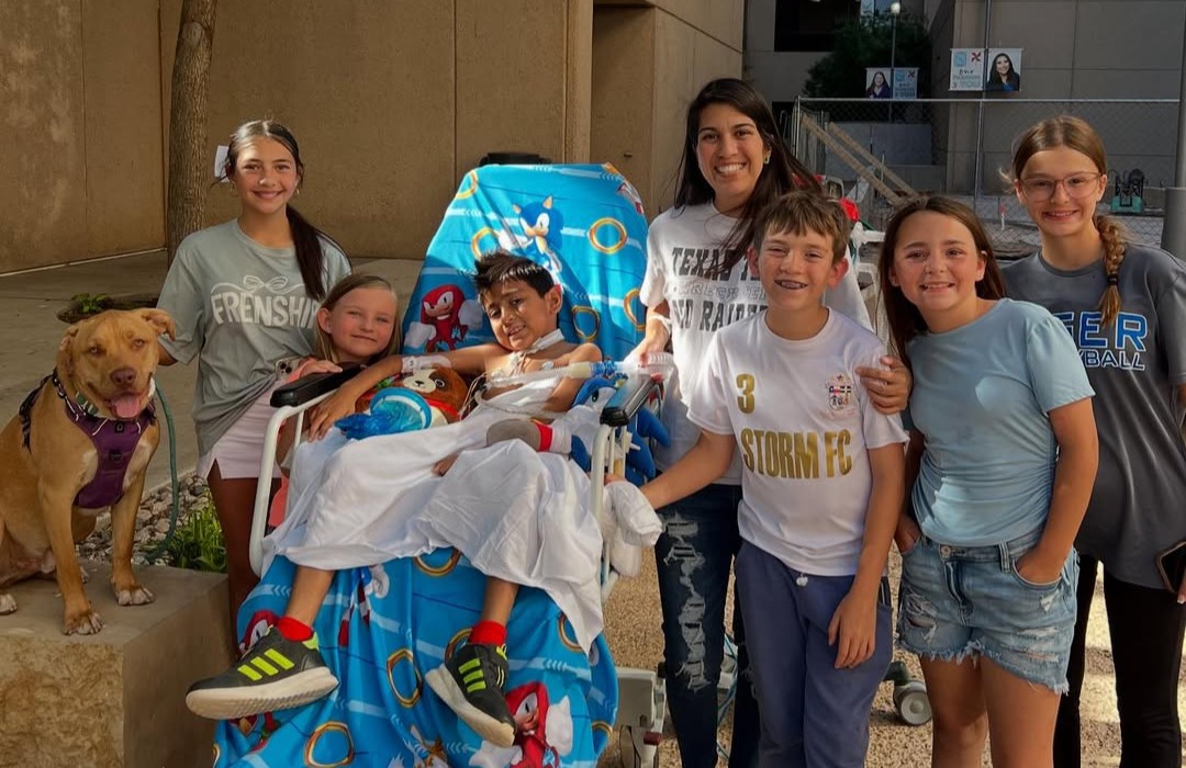
Featured image credit: Photo from Instagram — Casey Daniel (@caseyarend)
